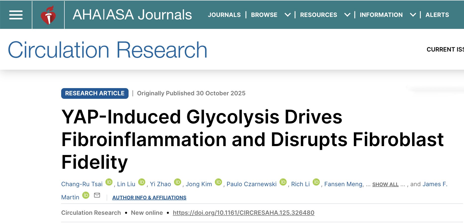
Dr. Tsai’s Cardiac Fibrosis Paper Published!
Our latest work reveals chamber-specific metabolic differences and their role in cardiac fibrosis
We're pleased to share our lab's latest paper, now published in Circulation Research. We're particularly proud that Dr. Chang-Ru Tsai's microscopy image was selected for the journal cover.
Chamber-Specific Differences in the Heart
Our study began with an unexpected observation: cardiac fibroblasts in the right atrium show higher YAP activity and increased glycolytic metabolism compared to fibroblasts in other cardiac chambers. This chamber-specific difference raised important questions about why these cells behave differently and what implications this might have for disease. The right atrium receives deoxygenated blood returning from the body, creating a relatively hypoxic environment. We hypothesized that this unique microenvironment contributes to the elevated glycolytic activity we observed.
YAP Activation Drives Glycolysis and Fibrosis
To understand YAP's role, we deleted the Hippo pathway kinases Lats1 and Lats2 that normally restrain YAP specifically in cardiac fibroblasts. The results were striking: the right atrium developed extensive fibrosis, inflammation, and arrhythmias, whereas the left atrium remained largely unaffected, despite having equivalent levels of Lats deletion. Our metabolic analyses revealed that YAP directly activates glycolytic genes, leading to increased glucose consumption and lactate production. Importantly, when we inhibited glycolysis or blocked lactate production, we could suppress the fibrotic response, demonstrating that glycolysis is functionally required for YAP-induced fibrosis.
Disruption of Fibroblast Identity
One of our most notable findings was that YAP activation causes adult cardiac fibroblasts to express SOX9 and acquire characteristics of osteochondroprogenitor cells. This represents a disruption of normal fibroblast lineage fidelity. We detected increased expression of chondrocyte and osteoblast markers, including accumulation of proteoglycans typically found in cartilage matrix. This finding suggests that cardiac fibroblasts have greater plasticity than previously recognized, with potential implications for understanding fibrotic remodeling.
Cell-Cell Communication Networks
Our single-cell and spatial transcriptomic analyses revealed important intercellular signaling mechanisms. YAP-activated fibroblasts secrete CSF1, which promotes macrophage expansion in the tissue. These macrophages, in turn, secrete IGF1, which signals back to the fibroblasts to enhance their proliferation and fibrotic activity. We validated these findings functionally: inhibiting CSF1 receptor signaling reduced macrophage numbers and suppressed fibroblast proliferation, osteochondroprogenitor differentiation, and fibrosis. Similarly, blocking IGF1 receptor signaling reduced fibrosis and fibroblast proliferation.
The cover image, captured by Dr. Tsai, shows cardiac tissue visualized through immunofluorescence microscopy. The image reveals the complex cellular architecture and distribution of different cell types within the fibrotic heart tissue, with multiple fluorescent markers highlighting specific cellular populations.
Translational Implications
These findings provide several insights relevant to cardiac fibrosis:
Chamber-specific susceptibility: The elevated baseline YAP and glycolytic activity in right atrial fibroblasts may explain why this chamber shows particular vulnerability to certain pathological changes.
Multiple intervention points: Our work identifies several potential therapeutic targets—glycolysis, lactate production, CSF1/macrophage signaling, and IGF1 signaling.
Metabolic-immune crosstalk: The reciprocal signaling between fibroblasts and macrophages highlights the importance of considering both metabolic and immune components in fibrotic disease.
Publication Details:
Title: YAP-Induced Glycolysis Drives Fibroinflammation and Disrupts Fibroblast Fidelity
Authors: Chang-Ru Tsai, Lin Liu, Yi Zhao, Jong Kim, Paulo Czarnewski, Rich Li, Fansen Meng, Mingjie Zheng, Jeffrey Steimle, Xiaolei Zhao, Francisco Grisanti, Zheng Sun, Jun Wang, Md. Abul Hassan Samee, Xiao Li, James F. Martin
Journal: Circulation Research, 2025
DOI: 10.1161/CIRCRESAHA.125.326480
Funding: This work was supported by the National Institutes of Health, American Heart Association, and Vivian L. Smith Foundation.
Key Findings:
Right atrial cardiac fibroblasts exhibit higher YAP activity and glycolytic metabolism than fibroblasts in other chambers
YAP directly activates glycolytic gene expression, and glycolysis is required for YAP-induced fibrosis
YAP activation disrupts fibroblast identity, inducing SOX9-expressing osteochondroprogenitor characteristics
YAP-activated fibroblasts and macrophages engage in reciprocal signaling through CSF1 and IGF1
Blocking either glycolysis, macrophage expansion, or IGF1 signaling reduces fibrosis


